2D Longitudinal Growth Study
When sequential cephalometric radiographs are available, we can track growth changes and treatment effects in the same individual through time. The unique historic role of growth studies is their ability to supply longitudinal data enabling us to track growth through time in individual persons.
1. The Craniofacial Growth Consortium Study (CGCS)
The Craniofacial Growth Consortium Study (CGCS) combines craniofacial records from six longitudinal growth studies (15,407 lateral cephalograms from 1,913 individuals; 956 females, 957 males, primarily European descent). Standard cephalometric points collected from the six studies in the CGCS allows direct comparison of craniofacial growth patterns across six North American locations.
The mission of the CGCS is to provide translational evidence-based outcomes to help shape critical research and clinical care of patients with craniofacial conditions.
The CGCS was conducted in both the CRIL and Dr. Richard Sherwood’s Craniofacial Growth Research lab at the University of Missouri, which was supported by the NIH (R01-DE024732). Dr. Oh is the Co-Principal Investigator of the grant.
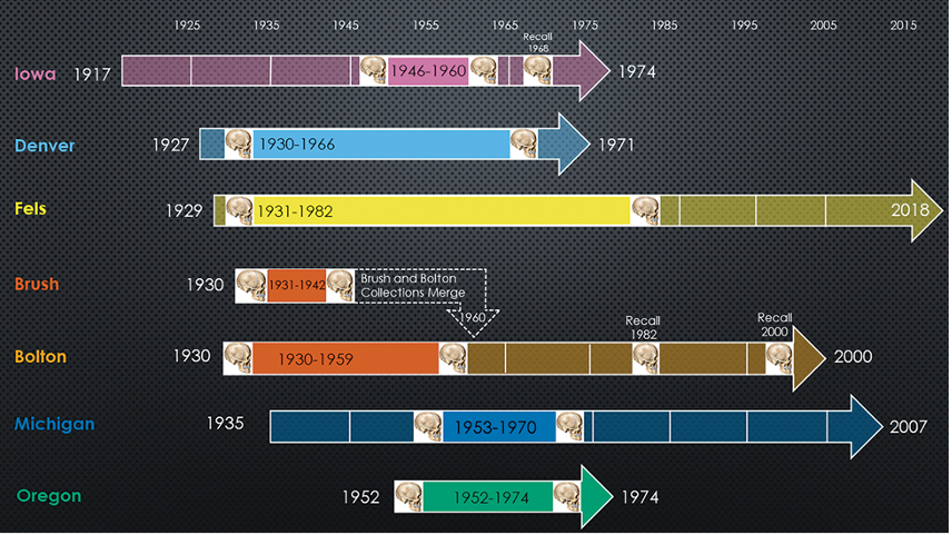
Below, timeline showing activity in each of the six growth studies comprising the CGCS. For each study, the period when cephalographs were collected is highlighted.
Ref. R Sherwood, H Oh, M Valiathan, K McNulty, D Duren, R Knigge, A Hardin, C Holzhauser, K Middleton. Bayesian Approach to Longitudinal Craniofacial Growth: The Craniofacial Growth Consortium Study. Anat. Rec. 2020; 1–29.
1) Mandibular plane angle changes from longitudinal series from ages 6 to 21
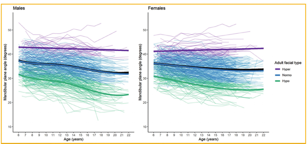
MPA decreased from childhood to adulthood in 92% of males and 81% of females, yet increased in 36% of males and 50% of females with the hyper-divergent adult facial type. Childhood MPA and overall change in MPA were significantly different by adult facial type.
Below, spaghetti plots of growth-related change in mandibular plane angle by adult facial type.
Ref. A Hardin, M Valiathan, H Oh, R Knigge, K McNulty, E Leary, D Duren, R Sherwood. Clinical implications of age-related change of the mandibular plane angle. Orthod Craniofac Res. 2020;23:50–58.
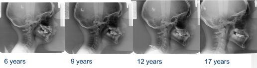
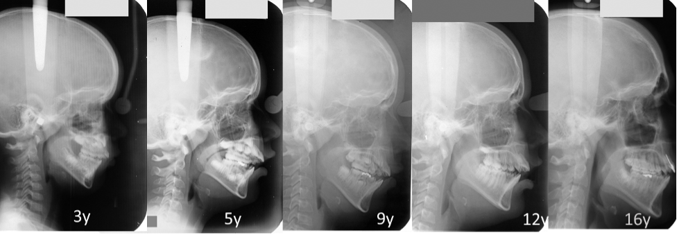
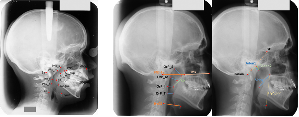
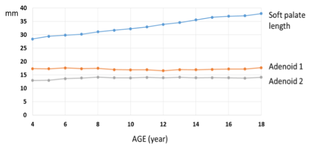
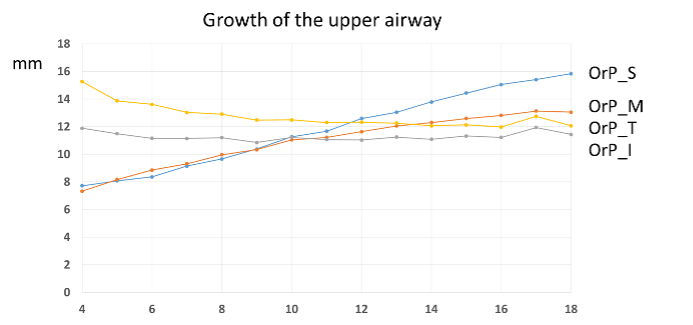
2) Longitudinal cephalometric study of Naso- and Oropharynx growth from 3 to 18 years
2. Geometric Morphometric Analysis
Understanding patterns of craniofacial growth is important for determining how to redirect or restrict growth to alter dental and skeletal relationships when treating patients. Identifying variations in integrated patterns of craniofacial growth that contribute to skeletal malocclusion has the ability to impact clinical approaches to treatment.
Geometric morphometric methods (GMM) was used to evaluate differences in craniofacial morphology and growth patterns among three skeletal facial types. The GMM approach allows for the evaluation of coordinated patterns of vertical and anteroposterior growth in craniofacial structures by preserving the geometric or spatial relationships of landmarks throughout the analysis, which traditional linear cephalometrics are poorly equipped to quantify.
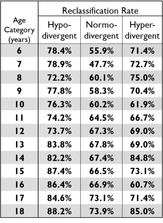
1) Predicting adult facial type from mandibular landmark data at young ages.
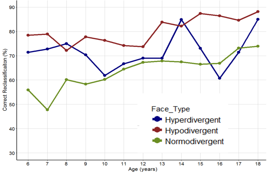
Ref. H Oh, R Knigge, A Hardin, R Sherwood, D Duren, M Valathan, E Leary, K McNulty. Predicting Adult Facial Type From Mandibular Landmark Data At Young Ages. Orthod Craniofac Res. 2019 May;22 Suppl 1:154-162.
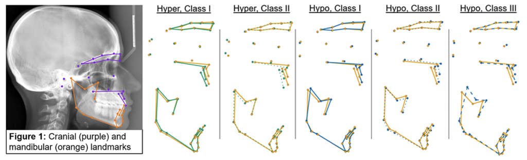
2) Geometric Morphometric Analysis of ontogenetic craniofacial covariation among facial classifications
Visualization of resulting morphology from each classification growth trajectory to 20 years of age, in comparison to Normo-divergent Class I (yellow, solid line configuration).
3) Geometric morphometric analysis of growth patterns among facial types
Below, differences between the average hyper-divergent (green) and hypo-divergent (blue) landmark configurations at ages 6, 10, 14, and 18.
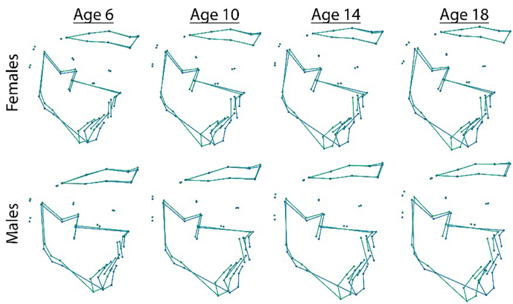
Ref: R Knigge, K McNulty, H Oh, A Hardin, E Leary, D Duren, M Valathan, R Sherwood. Geometric morphometric analysis of growth patterns among facial types. Am J Orthod Dentofacial Orthop 2020, August (Accepted).
3. The AAOF Craniofacial Growth Legacy Collection
The AAOF Craniofacial Growth Legacy Collection is the world’s first open access database of longitudinal records of growing adolescent subjects. The CRIL has been spearheading the AAOF Legacy Collection project, which has been supported by the AAOF (American Association of Orthodontists Foundation), since 2008.
CRIL has played a leading role in the construction of sharable electronic databases in orthodontics. Our laboratory web site has served as the prototype for a multi-institutional consortium sponsored by the American Association of Orthodontists Foundation which now developed, under our operational leadership, to sponsor the construction of a larger offshoot that will join representative samples from nine of the ten major longitudinal craniofacial growth collections in the U.S. and Canada. CRIL has been the central collection point and is building the consortium software platform based on its prior experience with or laboratory’s original site.
All cephalometric landmark data were collected in the CRIL. This Internet-accessible shared longitudinal records based of untreated subjects will mark a great advance for orthodontic teaching, research, and practice.
Go to the AAOF Legacy Collection website (www.aaoflegacycollection.org/aaof_home.html).
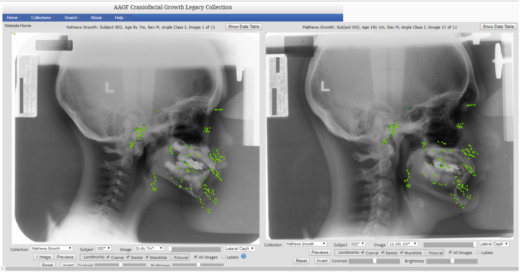
4. Mathews Implant Growth Study
The Mathews sample is the first long-term U.S. study of growing children with metallic implants of the Bjork type. The sample was collected by Dr. J. Rodney Mathews in the Section on Orthodontics, School of Dentistry, University of California San Francisco between the years of 1967 and 1979. Using open surgical methods, Dr. W. H. Ware, an associate of Dr. Mathews, placed 3 to 5 implants unilaterally in the mandible and a similar number arrayed transversely in the maxilla. Subjects were recalled at annual intervals between the ages of 7 and 18 years although few have records at more than 8 time points. Lateral Cephs, Posterior-Anterior Cephs, left and right 45-degree obliques were collected at each time point, but not all films have been digitized and traced at this time. Dr. Mathews’ original publication appeared in the Angle Orthodontist.1
Investigations of these records and data set at CRIL have resulted in a number of publications on dental and skeletal changes during preadolescent and adolescent growth.2-9 Among the most interesting observations from this data set has been the first documentation of systematic spontaneous post-natal widening of the core structures of the human mandible. 8,9 This observation has since been corroborated in two other longitudinal studies using subjects with implants.
References
- JR Mathews, GS Payne: Quantitative analysis of lower incisorchanges: A longitudinal implant study in man. Angle Orthod.50:218-29, 1980.
- S Baumrind, EL Korn, Y Ben-Bassat, EE West: The Quantitation of Maxillary Remodeling: 1. A description of osseous changes relative to superimposition on metallic implants. J. Orthod.91:29-45, 1987.
- S Baumrind, EL Korn, Y Ben-Bassat, EE West: The Quantitation of Maxillary Remodeling: 2. Masking of remodeling effects when an “Anatomical” method of superimposition is used in the absence of metallic implants. J. Orthod.91:463-474, 1987.
- S Baumrind, Y Ben-Bassat, EL Korn, LA Bravo and S Curry: Mandibular Remodeling Measured on Cephalograms: 1. Osseous changes relative to superimposition on metallic implants. J. Orthod. Dentof. Orthop.102:134-42, 1992.
- S Baumrind, Y Ben-Bassat, EL Korn, LA Bravo and S Curry: Mandibular Remodeling Measured of Cephalograms: 2. A comparison of information from implant and anatomical best fit superimpositions. J. Orthod. Dentof Orthop.102:227-38, 1992.
- S Baumrind, LA Bravo, Y Ben-Bassat, S Curry, EL Korn: Lower incisor and molar displacement associated with mandibular remodeling. Angle Orthodontist67: 93-102.
- S Baumrind, Y Ben-Bassat, LA Bravo, EL Korn, S Curry: Partitioning the Components of Tooth Displacement in the Maxilla by the Comparison of Data from Three Cephalometric Superimpositions. Angle Orthodontist66:117-130, 1996.
- EL Korn and S Baumrind: Transverse development of the human jaws between the ages of 8.5 and 15.5 years studied longitudinally with use of implants. Journal of Dental Research69:1298-1306, 1990.
- S Baumrind and EL Korn: Post-natal width changes in the internal structures of the human mandible: A longitudinal three-dimensional cephalometric study using implants. European Orthodontic Society14:417-426, 1992.
 Dr. J. Rodney Mathews (1911-1988) was a 1948 graduate of the University of California San Francisco’s “Curriculum II” specialty program in orthodontics. He had earlier pursued graduate studies in microbiology and had been involved in bacteriological research for the US Navy during World War II. At the UCSF School of Dentistry, studying under the mentorship of Wendell Wylie, his interest in the biology of growth and development deepened. For the next thirty-five years, Dr. Mathews divided his time between teaching and research at the UCSF Division of Orthodontics and Department of Growth and Development, and the private practice of orthodontics in Berkeley, California.
In his private orthodontic practice, he focused particularly on the early treatment of developing malocclusions, and in this area he collaborated closely with George Hahn. At the UCSF School of Dentistry, the main focus of his research was on the longitudinal study of human craniofacial growth with the aid of roengenographic cephalometrics.
Dr. Mathews was among the first investigators in the U.S. to recognize the seminal importance of Arne Bjork’s implant method to the study of craniofacial development. Between 1967 and 1985, he became the first and only American investigator to conduct a replication of the studies of Bjork for a sample of treated and untreated children observed longitudinally. Illness prevented his carrying these studies to completion but he graciously facilitated their continuation by CRIL. Through the continued kindness and support of the Mathews family, uniquely valuable images and data from these studies are being made available to the orthodontic specialty and the craniofacial research community on this web site.
Dr. Mathews was widely respected as a clinician and lecturer. He was featured in more than 100 presentations to local and national orthodontic groups, many in collaboration with his friend and colleague, Eugene E. West. As a teacher, he was always stimulating and patient. His publications were clear and concise. A hallmark of his presentations was his desire to stimulate information exchange among participants at all levels of academic sophistication.
Dr. Mathews was an early Diplomate of the American Board of Orthodontics and served as President of the Northern California Component of the Edward H. Angle Society. He was a member of Omicron Kappa Upsilon, the National Dental Honor Society, and of the Sigma Xi National Honorary Scientific Society.
Among his most widely known students are James A. McNamara, George S. Payne, Donald R. Poulton and Wayne Watson. His family continues his interest in craniofacial research by generous ongoing support of CRIL.