Data Science and Artificial Intelligence in Orthodontics
Data science includes data capture/acquisition, data processing, web-based storage and management, data analytics involving in-depth statistical analysis, machine learning (ML) approaches, and data communication. Artificial intelligence (AI) consists of developing computational systems that can perform human intelligence tasks, such as disease diagnosis, using features to help in the decision-making support.
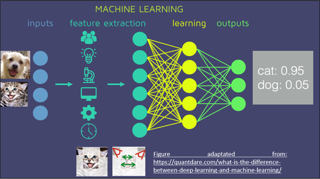
1. Artificial Intelligence and Machine Learning
Machine learning and artificial intelligence is widely used in other areas. In this example, a basic scheme shows which processes are involved in a basic machine learning algorithm.
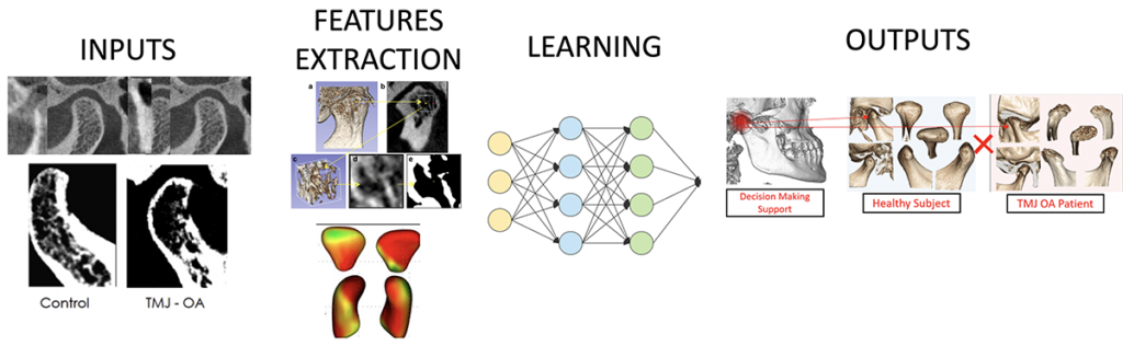
2. Artificial Intelligence and Machine Learning Applied to the TMJ Osteoarthritis Diagnosis
This figure illustrates the Inputs (several CBCTs images with TMJ OA or healthy); Features Extraction corresponds to the extraction of radiomics and shape features from the TMJ condyles; Learning is the machine learning algorithms model training; and Outputs shows the final prediction of the artificial intelligence model.
3. Data Science Applied in Orthodontics Research
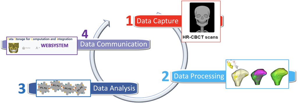
1. Data Capture
The 3D imaging research requires a standardized data science approach. First an acquisition of CBCT data (Cone-bean computed tomography exam) is acquired with different machines (left), and after acquisition, software to 3D reconstructs the image are necessary (right).
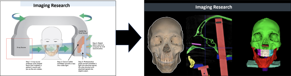
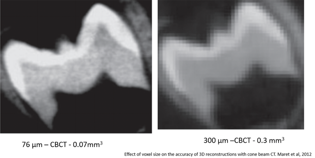
The voxel size is one of the parameters important to be investigated into the 3D research. Many measurement limitations can occur from non-adequate voxel size.
2. Data Processing
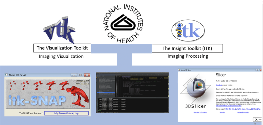
We use open-source software with pen libraries such as VTK and ITK, and 3D slicer to create our models and 3D imaging research procedures.
The next figure shows two distinct visualizations of the same CBCT image. In A, a 3D rendering was done. The 3D model is used only for visualization and cannot be used for surface measurement because it is not a real 3D surface model. In B, the CBCT image was segmented using the 3D slicer software (segmentation in green) and exported as the surface model (.stl or .vtk format); this 3D model is possible to place landmarks, planes and make measurements.
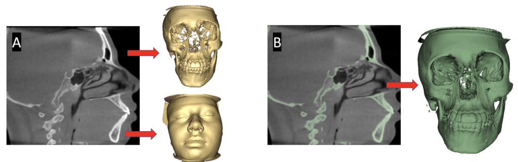
These videos illustrate the previous figure. Note that the segmented model (green) has more noise than the rendered model; however, it allows the user to refine the segmentation using semi-automatic software (ITK-SNAP) and the yellow only allows the visualization.
3. Data Analysis
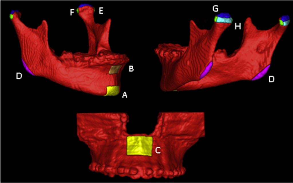
Examples of applications of 3D imaging measurement in the mandible region at different anatomical regions.
Bianchi, J., G. M. Porciúncula, L. Koerich, et al. “Three-Dimensional Stability Analysis of Maxillomandibular Advancement Surgery with and without Articular Disc Repositioning.” Journal of Cranio-Maxillofacial Surgery, vol. 46, no. 8, 2018, doi:10.1016/j.jcms.2018.05.031.