3D Imaging Research
Accurate measurement of three-dimensional changes in the craniofacial structures and the dentition is important. Using CBCT images we have developed 3D measurement tools for the diagnosis and evaluation of orthodontic treatment:
- 3D Landmark Reliability Study
- Integration of the Digital Study model to the CBCT Image
- 3D Facial Asymmetry Analysis
- Treatment Outcome Study
- Evaluation of Class II treatment outcome in CBCT
- 3D evaluation of naso- and oropharyngeal airway in various treatments
- Extraction vs. non-extraction
- Skeletal expansion (RPE, MARPE, DOME)
- 3D Evaluation of Condylar Joints in TMD Patients
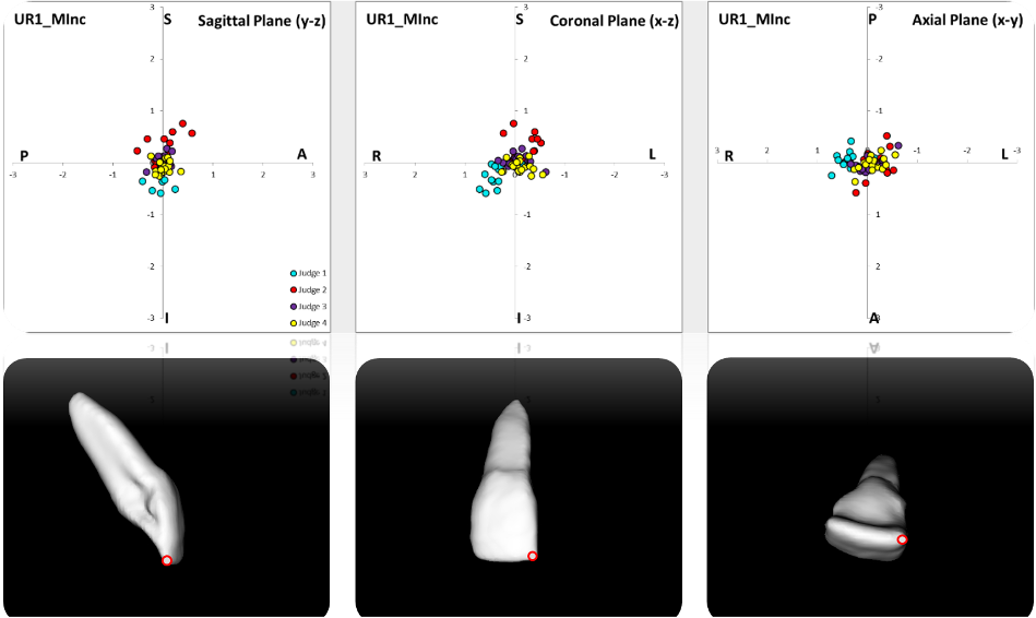
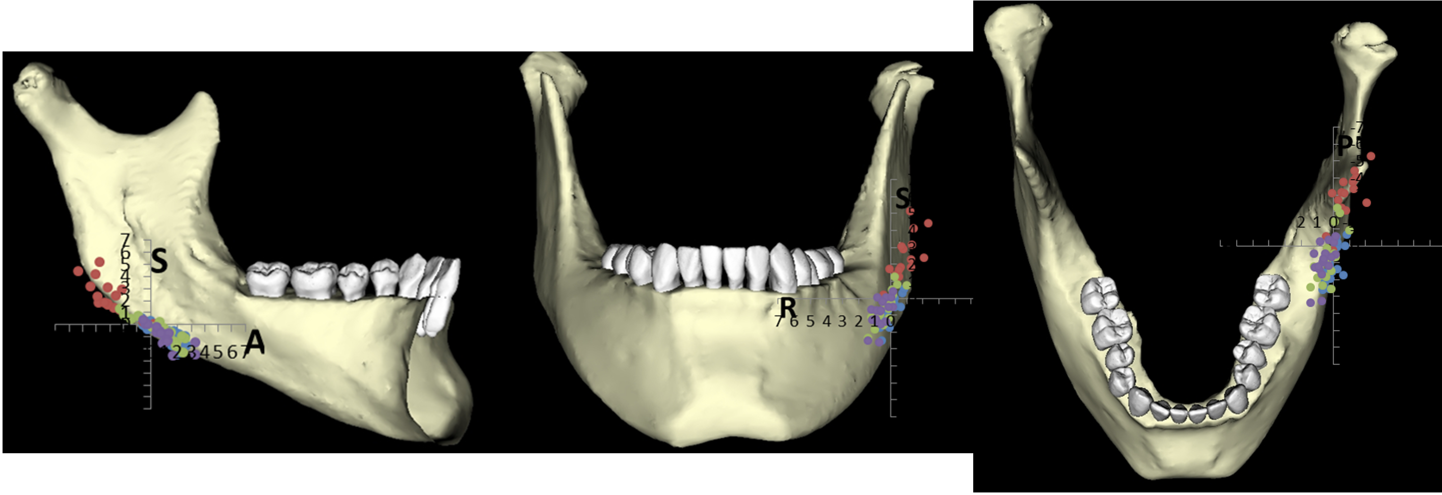
1. 3D Landmark Reliability Study
We quantified 3D reliability of skeletal and a comprehensive set of dental landmarks in CBCT to determine the shapes of envelope of error. There were differences in the size and shape of the distributions of errors of different landmarks. Most landmarks showed elongated envelopes. Bilateral structures tended to show greater errors than midline structures. Most dental landmarks were more reliable than skeletal landmarks.
Park J, Baumrind S, Curry S, Carlson S, Oh H, Boyd R. Reliability of 3D Dental and Skeletal Landmark Location on CBCT Images. Angle Orthod. 2019 Sep;89(5): 758-767
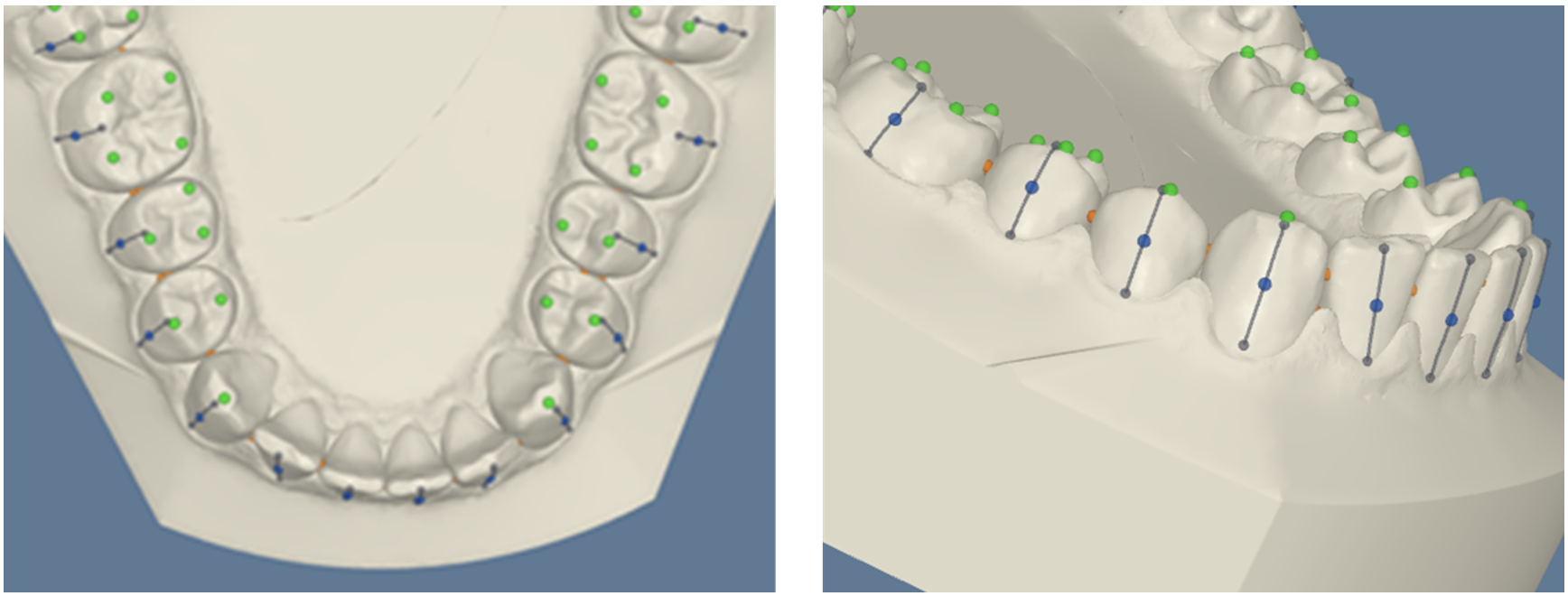
2. Integration of the Digital Study Model to the CBCT Image
Digital study models are by far the most important source of diagnostic information for orthodontists. They provide accurate 3D information of dentition for diagnosis of relative positions of the dentition. However, it does not provide any information in relationship to the maxilla, mandible, or the cranial base.
Digital study models obtained from intraoral scans (IOS) can be merged to the teeth on CBCT images. This type of integrated model provides accurate information of the dentition and its 3D relationship with the face.
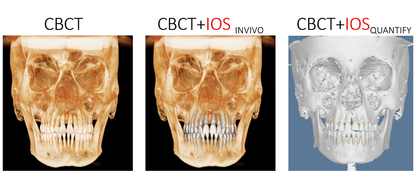
A. Quantify Project
Quantify is a software developed by Align Technology, and we have tested the accuracy of the tool which propagates dental landmarks from initial time point to the rest throughout treatment. The results of this study will be used to develop a tool which can track the position of individual tooth during the Invisalign treatment.
B. Accuracy of Integrating Digital Study Model to CBCT Image
We evaluated the accuracy of merging various types of IOS digital models.
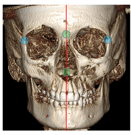
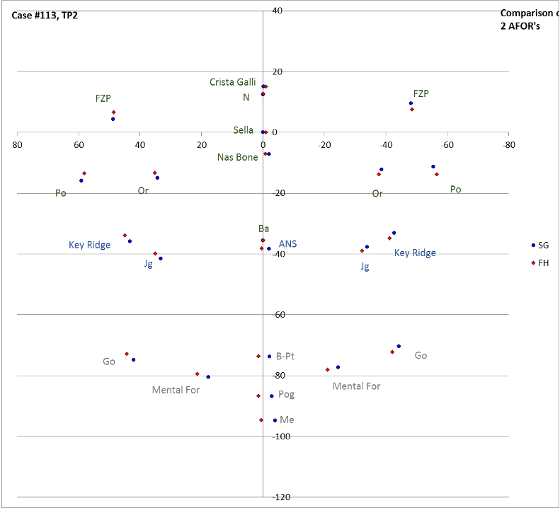
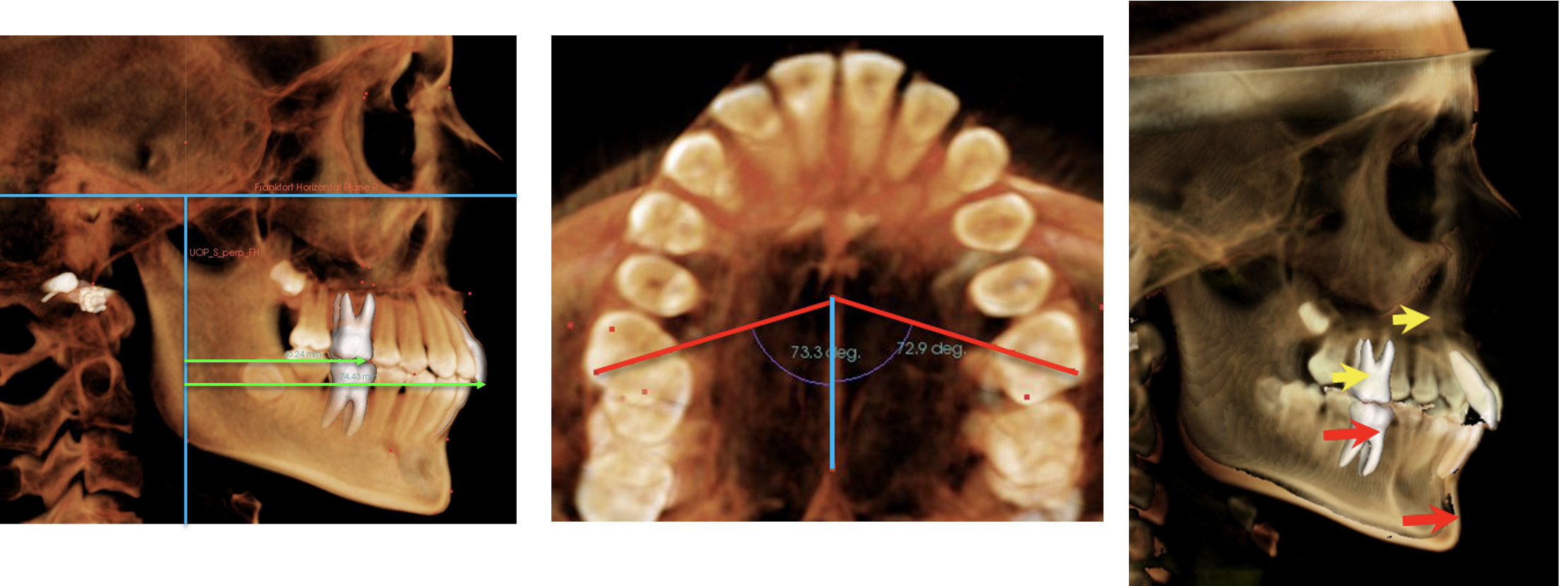
3. 3D Facial Asymmetry Analysis
Facial asymmetry can be quantified in a CBCT image. It’s evaluation heavily depends on the way the face is oriented which is based on the way the AFOR (Anatomical Frame of Reference) is constructed. We studied the effect of various types of AFOR and its effect on evaluation of facial asymmetry.
4. Treatment Outcome Study
We developed tools to measure various treatment outcome by using CBCT images in which 3D changes of positions of the jaws and dentition can be studied.
- Evaluation of Class II treatment outcome in CBCT
- 3D cephalometric characteristic of anterior openbite patients
- 3D evaluation of naso- and oropharyngeal airway in various treatments
- Extraction vs. non-extraction
- Skeletal expansion (RPE, MARPE, DOME)
A. Evaluation of Class II Treatment Outcome in CBCT
- We used CBCT images to identify the factors involved with Class II correction and to evaluate the treatment changes using 3D superimposition. 16 Class I patients (mean age 12.9 ± 1.3 years) and 22 Class II patients (mean age 12.5 ± 1.1 years) received orthodontic treatment. Voxel-based superimposition and landmark locations.
- Class II molar correction was achieved mostly through dentoalveolar effects with molar rotation being an important factor. Voxel-based superimposition is able to accurately and reproducibly superimpose 3-dimensional images. CBCT supersedes lateral cephalograms and study casts in analyzing orthodontic treatment effects.
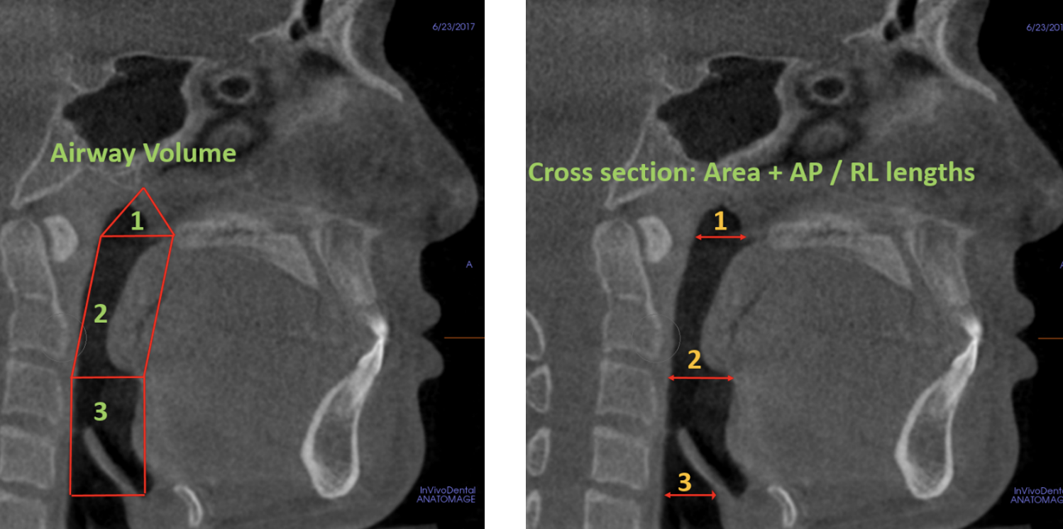
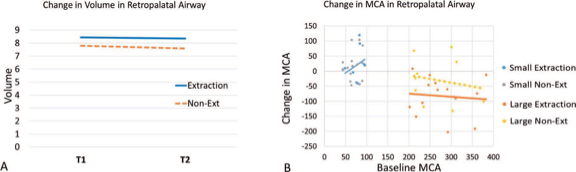
B. 3D Evaluation of Naso- and Oropharyngeal Airway in Various Treatments
- Airway and cephalometric changes in adult orthodontic patients after premolar extractions
- Joy A, Park J, Chambers DW, Oh H. Airway and cephalometric changes in adult orthodontic patients after premolar extractions. Angle Orthod. 2020 Jan;91(1):39-46.
- We examined changes in the airway and cephalometric measurements associated with orthodontic treatment of adults with and without premolar extractions. The study investigated whether extractions had a direct or indirect effect on the airway and examined selected and dental features.
- There was no evidence that extractions in nongrowing patients have negative consequences on the size of various airway measures in the nasopharynx, retropalatal, or retroglossal regions.
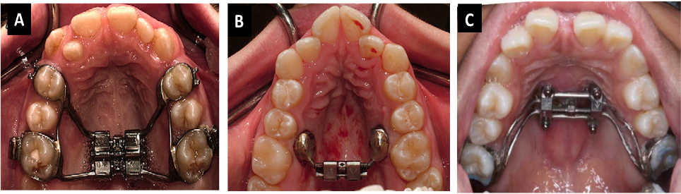
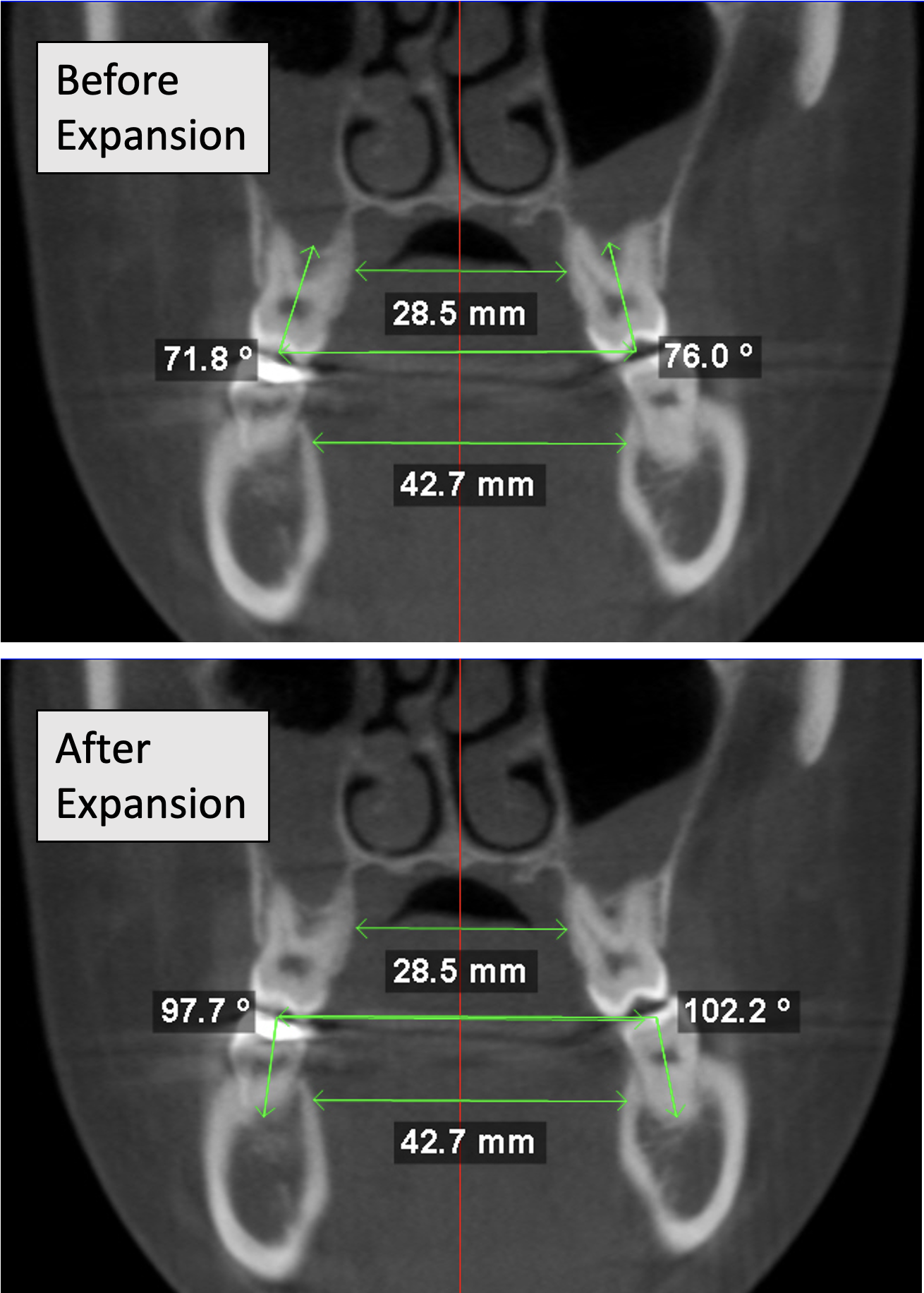
- Skeletal expansion (RPE, MARPE, DOME)
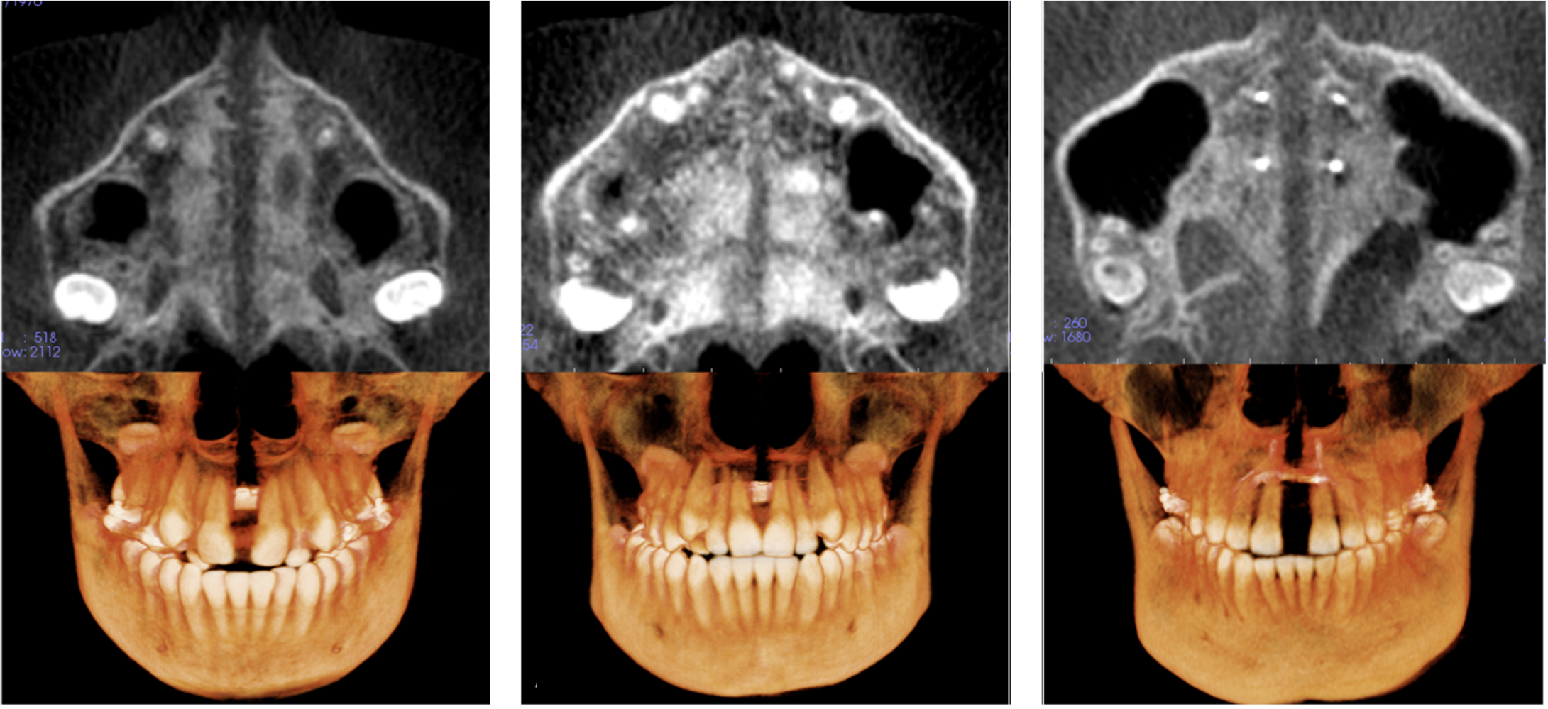
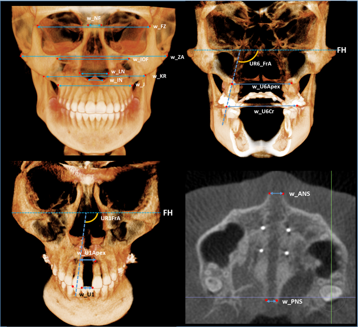
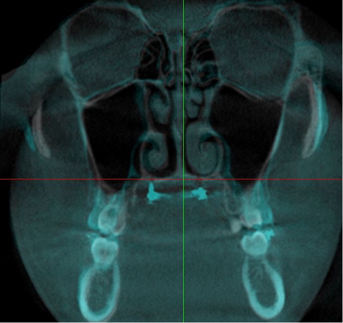
- Oh H, Park J, Lagravere-Vich M. Comparison of traditional RPE with two types of micro-implant assisted RPE: CBCT study. Sem in Orthod. 2019 Mar;25(1):60-68.
- Various types of the Micro-implant Assisted RPE (MARPE) are available to obtain greater skeletal expansion and to minimize dental effects. We evaluated skeletal and dental effects immediately after the completion of expansion using three different types of expanders — a traditional tooth-anchored maxillary expander (TAME) and two different types of MARPE, bone-anchored maxillary expander (BAME) and tooth-bone-anchored expander (MSE) using CBCT in adolescents.
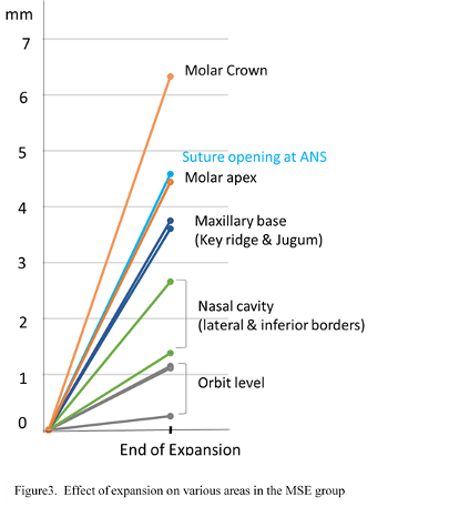
- Overall, the MSE group showed much greater skeletal changes than the TAME and BAME groups, especially, at the nasal floor, maxillary base, and palatal suture. About 72% – 78% of suture opening was at PNS, which indicates slightly more opening anteriorly than posteriorly; however, it was relatively parallel in nature than anticipated.
- In all three groups, the greatest transverse changes with expansion occurred at the molar crowns and the 2nd greatest changes at the palatal suture opening at ANS. It is suggested that MSE can be a great alternative method in correcting maxillary skeletal transverse deficiency.
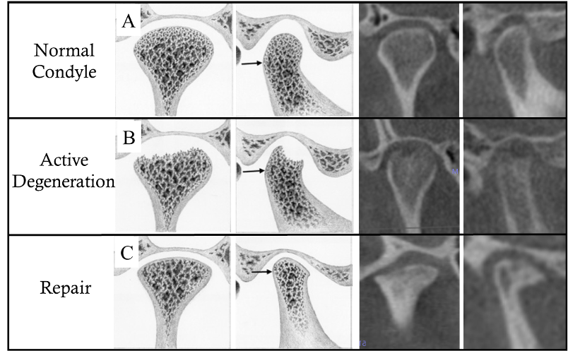
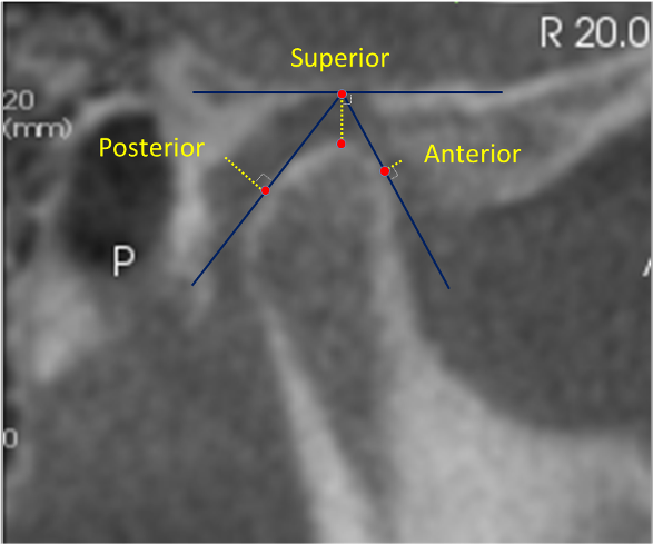
5. 3D Evaluation of Condylar Joints in TMD Patients
- Title: Condylar Degeneration in Anterior Open Bite Patients: A cone-beam computed tomography (CBCT) study
- Authors: Linda Phi; Brad Albertson; David Hatcher; Shikha Rathi; Joorok Park; Heesoo Oh
- Journal: Oral Surgery, Oral Medicine, Oral Pathology and Oral Radiology, 2021
- Objectives: The purpose of this study was to investigate the prevalence of condylar degeneration in anterior open bite patients.
- Study design: CBCTs of 194 anterior open bite patients and 100 control patients were included in this retrospective study. Two board-certified oral and maxillofacial radiologists reviewed the CBCT and categorized each condyle (588 condyles) as normal, degeneration-active, or degeneration-repair. Subjects with mandibular plane angles (MP-SN)≥38° were classified as skeletal open bites and those with less than MP-SN<38° as dental open bites. A chi-square test was used to evaluate the relationship between condylar status (normal vs. degeneration) and anterior open bites.
- Results: Of the 103 degenerative condyles, there were 59 condyles in the skeletal open bite group, 14 condyles in the dental open bite group, and 30 condyles in the control group. The Chisquare test indicated that condylar degeneration is associated with skeletal open bites, with condylar degeneration occurring twice as often in skeletal open bite patients as compared to the control (χ2=25.696, df=2, P<0.0001). Conversely, a higher rate of normal condyles was found in the dental open bite group (P=.0002).
- Conclusions: In skeletal open bite patients with high mandibular plane angles, it is important to suspect condylar degeneration as a potential etiology.